导读:来自厦门大学生命科学学院的研究人员在肝癌研究方面取得新进展,AIB1蛋白在68%的人肝癌标本中过度表达并可通过增强细胞增殖和侵袭能力而促进肝癌的进展。
科学家解析肝癌细胞侵袭机制
近期,来自厦门大学生命科学学院的研究人员在肝癌研究方面取得新进展,相关研究成果“Hepatitis B virus X protein stabilizes AIB1 protein and cooperates with it to promote human hepatocellular carcinoma cell invasiveness” 于4月2日发表在国际期刊《肝脏病学》(Hepatology)(影响因子10.885)上。
文章通讯作者是厦门大学生命科学学院俞春东教授,其课题组主要从事核受体及其共激活子在肝脏代谢及肝脏疾病中的作用,及核受体及其共激活子在炎症反应及自身免疫疾病中的作用等研究。论文的第一作者为09级硕士生刘永宏。
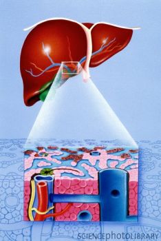
人类肝细胞癌(HCC)是最常见的肝脏肿瘤,在世界范围内肿瘤相关性死亡因素中排名第三,全球每年有超过50万新患者,中国新病例占了全球所有肝癌病例的一半以上(55%)。目前临床上最主要治疗肝癌的方法是手术切除和肝脏移植,经肝动脉栓塞的化疗法则作为第二线治疗。但是由于肝癌复发率高、且抗化疗药性强,整体治疗效果并不理想。多年来科学家们一直致力于解析肝癌发生发展机制以寻找有效的预后判断标志物与治疗靶标。
乙型肝炎病毒(HBV)慢性感染与人类肝细胞癌的发展密切相关,而乙肝病毒X蛋白(HBx)在肝癌的进展中起着关键的作用。AIB1蛋白在68%的人肝癌标本中过度表达并可通过增强细胞增殖和侵袭能力而促进肝癌的进展。由于HBx和AIB1在肝癌进展中发挥着重要的作用,研究人员希望了解它们是否协同促进了肝癌的发展。
在这篇文章中,研究人员发现与HBx阴性肝癌组织相比,HBx阳性肝癌组织中AIB1蛋白的水平较高,并且HBx蛋白水平和AIB1蛋白水平呈正相关。在不影响其mRNA水平的情况下,HBx可通过延长AIB1蛋白的半衰期来使AIB1的蛋白水平增加。HBx蛋白通过与AIB1的结合来阻止E3 泛素连接酶Fbw7a和AIB1的结合,进而抑制Fbw7a对AIB1的泛素化降解。此外,HBx和AIB1可募集到MMP-9 启动子上协同促进MMP-9启动子的活性,进而增强MMP-9在HepG2 细胞中的表达而促进细胞的侵袭能力。
该研究表明HBx可稳定AIB1蛋白并与它协同促进肝癌细胞的侵袭,说明HBx和AIB1之间的相互作用在乙肝病毒相关的肝癌进展中起着重要的作用。

 Hepatitis B virus X protein stabilizes AIB1 protein and cooperates with it to promote human hepatocellular carcinoma cell invasiveness
Liu, Yonghong; Tong, Zhangwei; Li, Ting; Chen, Qiang; Zhuo, Luting; Li, Wengang; Wu, Ray‐Chang; Yu, Chundong
Chronic infection of Hepatitis B virus (HBV) is closely associated with the development of human hepatocellular carcinoma (HCC). HBV X protein (HBx) plays a key role in the progression of HCC. We recently found that amplified in breast cancer 1 (AIB1) protein is over-expressed in 68% human HCC specimens and promotes HCC progression by enhancing cell proliferation and invasiveness. Given that both HBx and AIB1 play important oncogenic roles in HCC, we aim to determine whether they can cooperatively promote human HCC development. Herein we showed that HBx-positive HCC tissues had higher level of AIB1 protein compared to HBx-negative HCC tissues. A positive correlation between HBx protein level and AIB1 protein level was established in HCC specimens. Without affecting its mRNA level, HBx induced a significant increase of the protein level of AIB1, which correlated with a significant extension of the half-life of AIB1 protein. Mechanistically, HBx could interact with AIB1 to prevent the interaction between E3 ubiquitin ligase Fbw7 and AIB1, and then inhibited Fbw7-mediated ubiquitination and degradation of AIB1. In addition, reporter assays and ChIP assays revealed that both HBx and AIB1 were recruited to MMP-9 promoter to enhance MMP-9 promoter activity cooperatively. Consistently, HBx and AIB1 cooperatively enhanced MMP-9 expression in HepG2 cells, which in turn increased cell invasive ability. Conclusion: Our study demonstrates that HBx can stabilize AIB1 protein and cooperate with it to promote human HCC cell invasiveness, highlighting the essential role of the crosstalk between HBx and AIB1 in HBV-related HCC progression.
文献链接:https://onlinelibrary.wiley.com/doi/10.1002/hep.25751/abstract


